Causes of tethered cord syndrome
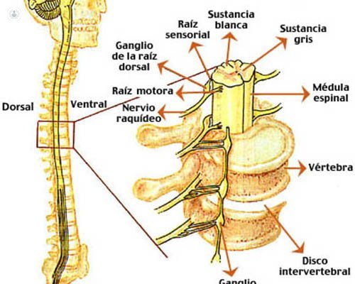
Written by:Under normal conditions the spinal cord is subject within the spinal canal by ligaments and jagged terminal phylum. The latter is a band of fibrous connective tissue that allows some movement of the conus to small movements of flexion and extension. Thus, the fixed terminal phylum and stabilizes the spinal cord while absorbing impacts and excessive movements thereon.
If there is any reason to limit the mobility of the spine (for example, fatty infiltration of the phylum or lipomas conus medullaris that can produce traction and tension stretching in flexion and extension of the spine) abnormal stress may occur on the cone (the end of the spinal cord) that would lead to tethered cord syndrome ( "tethered cord syndrome").
Causes of tethered cord
There are two hypotheses that attempt to explain the neurological deterioration that may occur in a central cone anchored. On the one hand, the mechanical stress caused by traction on the bone of the elements that anchor, whether a lipoma, a terminal phylum or thickened bands fibroconectivas. Furthermore, ischemia caused by blood flow defects caused by stress on the core microvasculature.
Variability in the circulation of cerebrospinal fluid (CSF) that bathes and protects the spinal cord, and other individual factors such as vascularization, the size of the spinal cord and its position on the channel or the anchor point, could determine differences in the Similar symptoms among patients.
In most cases, tethered cord is secondary to congenital malformation produces an "anchor", an impossibility of movement of the spinal cord within the vertebral bone tunnel, thereby resulting in microtraumatismos and defects blood supply stretch vessels that nourish the spinal cord. This deficit, if sustained over time, can lead eventually to a spinal cord injury.
The most common causes of congenital malformations that produce a tethered cord lipomas are the core and dermal sinuses, although there are a variety of them across the spectrum of occult spina bifida (diastematomielia, "dorsal limited myeloschisis", etc.).
Some patients operated tethered cord may develop late and after the first intervention a new anchor ( "re-anchor"), produced by an internal fibrous scar. It is the second most common cause of tethered cord operation, but fortunately not a common complication.
Neurosurgery for tethered cord
In cases of spina bifida occurring with hidden tethered cord, lipoma as the medullary cone, dermal sinus or diastematomielia, treatment should be surgical if symptoms occur derived therefrom. In some cases preventive surgical treatment is recommended even in asymptomatic patients, to prevent future complications that might result from a spinal cord injury caused by the anchor.
Surgical treatment of spina bifida and tethered cord hidden generally has a good prognosis. But both the indication for surgery, as its potential risks and benefits should be discussed with the patient and / or family members based on each case.
The goal of surgery is to prevent neurological deterioration or progression if they already existed prior symptoms. As a general rule must be operated all symptomatic cases or cases intervened before presenting new symptoms or progression of the previous deficit ( "re-anchor").
The general principles include resection of the mass of the lipoma, the full anchoring of the spinal cord, neurological tissue preservation, and restoration of the anatomical planes (including the tubulation of the neural plate and repairing their roofs).
The indication for surgery in the case of lipomas should take into account two variables: the type of lipoma, and if this is symptomatic or not. There are more favorable than others to get a resection with disembedding without the appearance of complications cases. In this way, it is easier to make the decision to indicate surgery in patients with terminal lipoma flow or phylum than in those with a complex lipoma.
The experience of the neurosurgeon in these cases has a major influence on achieving satisfactory results.