Tired of suffering watery eyes?
Written by:Tearing is an important part of the consultations that will cater services in Ophthalmology. Although it may seem a trivial disease, it is not for the sufferer and, while not generating serious health implications of the individual, can become extremely disabling for the patient, with significant psychological and occupational consequences.
Structure of the tear ducts
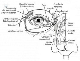 The tear ducts comprise the tear lake, located on the surface of the eye; the puncta, where tears fall and subsequently run through the upper and lower lacrimal canaliculi that empty into the lacrimal sac. This tapering and forming the nasolacrimal duct, which runs the bone and ends at the nostril. A failure anywhere along this pathway may delay or block the drainage of tears and cause tearful eye.
Crying eyes
Sometimes watery eyes accompanied by signs of infection such as conjunctivitis repeat, discharge from the eye lashes and even dacryocystitis, which are serious infections that generate large lacrimal sac inflammation, severe pain and malaise.
Diagnosis and treatment of diseases of the tear ducts
For the location of the tear obstruction will do some diagnostic tests: probing and irrigation of the tear duct with a cannula, instillation of drops of dye into the eye and observing their way into the nose and can even perform radiological tests show us how the contrast.
If a tear duct pathology that justifies tearing, treatment depends on the location of the blockage. Usually we perform a surgical treatment that we can practice under local anesthesia and on an outpatient basis. Occasionally, the puncta are those closed or very narrow and do not allow the entry of the tear in the circuit. In this case the treatment will puntoplastia, ie small cuts in the puncta to expand the hole where the tear enters the tubules.
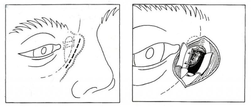 When the obstruction is lower, we resort to dacryocystorhinostomy, which is internally join the lacrimal sac to the lining inside the nose, creating a new form of tear drainage. The classic approach to this intervention is the external, from the skin, so called external dacryocystorhinostomy. In recent years there has been innovation in this field, appearing minimally invasive techniques, which can perform surgery from the nose or by laser from within the lacrimal canaliculus, thereby disappears wound skin and is greatly reduced surgical trauma.
In cases where conventional techniques fail or obstruction is likely to be treated by them, one resorts to the implantation of a glass tube that connects the inside of the lacrimal lake or wattle nose, creating a direct communication for draining the tear, the lacorrinostomía.